
In the field of orthopedics, the clavicle often emerges as the most frequently fractured bone, attributed to its slender structure and superficial location. This bone, commonly referred to as the collarbone, plays an essential role in shoulder mechanics yet is highly susceptible to fractures. Understanding why the clavicle is particularly prone to breaking involves examining its anatomical features and the common mechanisms of injury. Such insights are vital for both healthcare professionals and individuals engaged in high-impact activities. What makes the collarbone's vulnerability a focal point in discussions about bone fractures?
Understanding Bone Fractures
How do bone fractures occur, and what factors contribute to their susceptibility? Bone fractures transpire when a force exceeding the bone's inherent strength is applied, leading to a break in the continuity of the bone tissue. The arm, particularly the humerus, is frequently prone to fractures resulting from falls or direct impacts, common in both pediatric and adult populations. Similarly, wrist fractures, especially those involving the distal radius, are prevalent in younger individuals engaging in high-risk activities such as skiing or contact sports.
The anatomical structure and mechanical function of bones play a pivotal role in their susceptibility to fractures. For instance, the clavicle, due to its superficial positioning and the forces it absorbs during shoulder impacts, is notably susceptible. In the lower extremities, the ankle bones, comprising the tibia, fibula, and talus, are vulnerable to fractures from high-impact activities like running or sudden directional changes. These fractures are often seen in athletic populations and result from the substantial forces transmitted through the joint. Understanding the specific mechanics and vulnerabilities of different bones provides significant insights into their propensity for fractures, emphasizing the need for targeted preventive strategies in at-risk activities and populations.
Why Bones Break
Bones fracture primarily due to significant impact or trauma from incidents such as falls, vehicular collisions, or athletic activities. Additionally, bones compromised by conditions like osteoporosis exhibit reduced structural integrity, markedly increasing susceptibility to fractures. Understanding these mechanisms is essential for the development of effective prevention strategies and therapeutic interventions.
Impact and Trauma
Understanding the mechanisms of impact and trauma elucidates why certain bones are more susceptible to fractures. Clavicle fractures occur frequently due to the clavicle's anatomical position and its role in bearing the brunt of falls or direct blows to the shoulder. This bone's relative thinness and superficial location make it particularly vulnerable.
Arm bones, including the humerus and ulna, are also prone to fractures from falls or high-impact injuries. The humerus, being the long bone of the upper arm, can sustain fractures near the shoulder, mid-arm, or elbow, depending on the nature of the trauma. The ulna, one of the two long bones in the forearm, often fractures alongside its counterpart, the radius, especially when an individual instinctively uses their arm to break a fall.
Wrist fractures are common, specifically involving the distal radius, also known as a Colles fracture. Such fractures frequently occur in younger individuals engaged in high-risk activities like skiing, where the impact forces are transmitted through the wrist upon falling.
Bone Weakness Factors
Numerous intrinsic and extrinsic factors contribute to bone weakness, greatly increasing the risk of fractures. Osteoporosis, a condition characterized by decreased bone density and structural deterioration of bone tissue, is a primary intrinsic factor. It greatly elevates fracture risk, particularly in the elderly population. Poor bone density, often a result of nutritional deficiencies such as inadequate calcium and vitamin D intake, further exacerbates susceptibility to fractures.
Extrinsic factors also play an essential role in compromising underlying bone health. Medications like corticosteroids can precipitate bone resorption, while hormonal imbalances, particularly reduced estrogen levels in postmenopausal women, intensify bone demineralization. Lifestyle choices such as physical inactivity, smoking, and excessive alcohol consumption contribute to bone fragility by impairing bone remodeling processes.
Genetic predispositions can also influence bone strength, predisposing individuals to conditions that weaken bones. Environmental factors, including falls, accidents, and sudden impacts, can easily fracture weakened bones, especially in individuals with compromised bone health.
Preventive measures, such as regular bone density screenings and lifestyle modifications—like increased physical activity and dietary adjustments—are essential for maintaining bone health and minimizing fracture risk. Addressing these factors is critical for reducing the incidence of fractures and improving overall skeletal integrity.
Anatomy of the Collarbone
The clavicle, a slender S-shaped bone, serves as a strut between the sternum and scapula, playing a pivotal role in shoulder mechanics and upper limb mobility. This bone, commonly referred to as the collarbone, is situated proximally near the front side of the chest, adjacent to the shoulders. Its anatomical position renders it susceptible to fractures, particularly in children, where it represents a significant portion of skeletal injuries. Anatomically, the clavicle serves as a critical component in maintaining the structural integrity and alignment of the shoulder girdle.
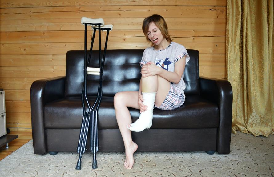
When a collarbone fracture occurs, it typically results from direct trauma, such as a fall onto an outstretched arm or a forceful impact to the shoulder. The healing process entails a multifaceted approach, often beginning with immobilization to ensure proper bone alignment and stabilization. This is typically achieved through the use of slings or figure-of-eight bandages. Subsequently, physical therapy becomes indispensable in the rehabilitation phase, aiming to restore strength, flexibility, and function to the affected shoulder. In cases of severe clavicle fractures, surgical intervention may be necessary to realign and stabilize the bone fragments, facilitating the best possible healing and preventing long-term complications.
Common Causes of Fractures
Common causes of bone fractures include accidental falls, which are particularly prevalent among the elderly due to osteoporosis-induced bone fragility. High-impact sports and physical activities, such as skiing and running, frequently result in fractures due to sudden twists or collisions. Additionally, osteoporosis compromises bone strength, making fractures more likely even from minor stresses or impacts.
Accidental Falls Impact
Accidental falls frequently result in fractures of the clavicle, a bone particularly vulnerable due to its anatomical position and structural characteristics. Clavicle fractures are prevalent because the clavicle, or collarbone, serves as a strut between the sternum and the scapula, making it susceptible to the impact of falls. This is especially true in children, where the clavicle is one of the most commonly fractured bones owing to its relatively thin and elongated shape.
The mechanism of injury typically involves a direct impact to the shoulder area during a fall, which transmits force along the clavicle, leading to a fracture. Management of a clavicle fracture often necessitates shoulder immobilization using a sling or figure-eight brace to maintain proper alignment during the healing process. This immobilization helps in reducing pain and preventing further displacement of the bone fragments.
Following the initial immobilization phase, physical therapy plays a significant role in recovery. Physical therapy is designed to restore range of motion, strengthen the surrounding musculature, and promote functional recovery of the shoulder joint. The combination of shoulder immobilization and physical therapy is essential for best healing and to prevent long-term complications associated with clavicle fractures resulting from accidental falls.
Sports-Related Injuries
Many sports-related injuries, particularly those involving high-impact activities such as football, hockey, and cycling, frequently result in clavicle fractures. The clavicle, or collarbone, is significantly susceptible to fractures due to its anatomical position and structural characteristics. In these sports, athletes are often exposed to significant impact or falls, which can easily lead to a broken clavicle.
The high incidence of clavicle fractures in sports-related injuries is attributed to the direct impact forces and the nature of physical contact inherent in these activities. For example, in football and hockey, players are at risk of collisions that exert substantial force on the upper body, making the clavicle vulnerable. Similarly, cyclists who experience falls often attempt to catch themselves with an outstretched arm, transmitting force through the clavicle.
Preventive measures, including the use of appropriate protective gear and adherence to proper training techniques, are critical in mitigating the risk of clavicle fractures. Protective gear such as padded vests and shoulder pads can absorb some of the impact forces, thereby reducing the likelihood of fracture. Additionally, educating athletes on safe playing techniques can further decrease the incidence of these injuries.
Osteoporosis and Weak Bones
Osteoporosis, a prevalent metabolic bone disease characterized by decreased bone mass and deteriorated bone tissue, markedly heightens the risk of fractures in affected individuals. This condition is particularly common among older adults, with women being disproportionately affected due to hormonal changes post-menopause that accelerate bone density loss. The intrinsic fragility of osteoporotic bones renders them susceptible to fractures even under minimal trauma or falls, which are often unavoidable in this demographic.
Low bone density, a hallmark of osteoporosis, greatly compromises the structural integrity of bones, leading to weak bones that are prone to fractures. These fractures frequently occur in the wrist, hip, and spine, areas that bear substantial mechanical stress. The clinical consequences of such fractures can be severe, often leading to reduced mobility, chronic pain, and in some cases, increased mortality.
Preventive measures are paramount in mitigating the risk of osteoporosis-related fractures. Adequate calcium and vitamin D intake, weight-bearing exercises, and pharmacologic interventions such as bisphosphonates or selective estrogen receptor modulators can enhance bone density and strength. Early diagnosis and proactive management of osteoporosis are critical in preserving bone health and preventing debilitating fractures.
Symptoms of a Broken Collarbone
Common symptoms of a broken collarbone include acute pain, localized swelling, and marked tenderness at the fracture site. These symptoms often manifest immediately following the injury, making early identification essential. The pain associated with a fractured collarbone can be intense, exacerbated by movement of the shoulder or arm. Swelling typically develops rapidly around the fracture area, contributing to discomfort and visible deformity.
A notable deformity or palpable bump at the site of the fracture is a hallmark sign, indicative of the bone's misalignment. Such deformities can vary in prominence but generally suggest a significant structural compromise. In addition to these primary symptoms, limited range of motion in the shoulder or arm is common. Patients often report a reluctance or inability to move the affected limb due to severe pain.
Nerve involvement, though less common, can present as numbness or tingling in the arm or shoulder area, suggesting possible nerve compression or injury. Prompt medical evaluation is imperative to confirm the diagnosis and initiate appropriate treatment. Ignoring these symptoms can lead to complications, emphasizing the importance of early medical intervention for a suspected broken collarbone.
Diagnosing a Fractured Collarbone
Thoroughly diagnosing a fractured collarbone involves a detailed clinical evaluation, often supplemented by imaging techniques such as X-rays to confirm the presence and extent of the fracture. The diagnostic process begins with a detailed physical examination, where the clinician inspects for visible deformities, swelling, and tenderness along the clavicle. Palpation helps identify the precise location of the fracture and assess any associated soft tissue injury.
Imaging tests are indispensable in diagnosing a fractured collarbone. Standard anterior-posterior and cephalic tilt X-rays provide clear views of the clavicle, revealing fracture lines, displacement, and angulation. In certain cases, advanced imaging modalities such as computed tomography (CT) scans may be employed for a more thorough assessment, especially when the fracture is complex or involves intra-articular extension.
Timely and accurate diagnosing is crucial for the proper management of a fractured collarbone. Early intervention allows for appropriate treatment planning and helps mitigate complications, such as malunion or nonunion. Prompt medical attention ensures that the fracture is managed effectively, facilitating efficient healing and functional recovery.
Treatment Options
Treatment options for fractures, including those of the collarbone, encompass immediate first aid measures and subsequent medical interventions. Immediate first aid typically involves immobilization and pain management, while medical interventions may range from casting and splinting to surgical fixation for more complex or displaced fractures. Ensuring proper rehabilitation and follow-up care is imperative for best possible recovery and functional restoration.
Immediate First Aid
Immediate first aid for a fractured bone necessitates vital immobilization of the affected area to prevent further injury and facilitate ideal healing conditions. Immobilizing the injured limb is essential and can be achieved using a splint or any rigid material available to stabilize the bone. This helps to minimize movement and reduce the risk of exacerbating the fracture.
Elevating the affected limb above the level of the heart is recommended to decrease swelling and alleviate pain. Elevation aids in reducing fluid accumulation at the injury site, which can contribute to discomfort and prolonged healing times.
Applying ice to the injury site within the first 48 hours can greatly reduce inflammation and numb the area, providing symptomatic relief. Ice should be applied intermittently, typically in 20-minute intervals, to avoid frostbite.
It is imperative to seek medical attention promptly to guarantee accurate diagnosis and appropriate treatment. Professional medical evaluation is necessary to determine the extent of the fracture and to formulate a detailed treatment plan. Avoid excessive movement of the injured limb to prevent worsening the condition. Immediate first aid measures are essential to optimize outcomes before definitive medical care is administered.
Medical Interventions
Management of a clavicle fracture typically involves conservative measures such as immobilization with a sling, although severe cases may require surgical intervention to guarantee proper alignment and healing. This common fracture, especially prevalent in children due to their active lifestyles and the slender structure of the clavicle, is generally approached first with non-operative treatment. Immobilization with a sling provides the necessary support, allowing the bone to heal naturally over several weeks.
However, certain conditions that cause bones to break more easily, such as osteoporosis or significant trauma, may complicate the fracture. In these instances, fractures often require surgery to ensure the correct treatment and alignment of the bone. Surgical intervention typically involves the use of plates, screws, or rods to stabilize the fracture and promote proper healing. This approach is particularly vital in cases where the bone is significantly displaced or fragmented.
Post-surgical care includes physical therapy to restore strength and mobility, often resulting in a quicker return to normal function compared to conservative methods. Understanding the correct treatment for this common broken bone is essential for the best recovery and prevention of long-term complications.
Healing and Recovery
Healing a fractured clavicle generally necessitates immobilization with a sling or brace to facilitate proper bone alignment and natural recovery. This immobilization is important as it prevents unnecessary movement, allowing the bone to heal in the correct anatomical position. Typically, a sling or brace is recommended for a duration of 4-6 weeks, depending on the severity of the fracture and the patient's overall health status.
Recovery from a clavicle fracture extends beyond initial immobilization. The complete healing process generally spans 6-8 weeks, during which the patient must adhere to medical advice, avoiding strenuous activities that could compromise the integrity of the healing bone. Upon confirmation of adequate bone healing through radiographic imaging, physical therapy is often initiated to restore shoulder strength and range of motion.
Physical therapy plays an important role in the rehabilitation phase, addressing potential muscle atrophy and joint stiffness developed during immobilization. Therapeutic exercises are designed to gradually increase flexibility, strength, and functional capacity of the shoulder girdle. Adherence to a structured physical therapy regimen, combined with proper rest and gradual reintroduction of activities, greatly enhances the likelihood of a full and timely recovery.
Preventing Collarbone Fractures
Preventing collarbone fractures necessitates a multifaceted approach that includes proper protective gear, strength training, and awareness of risk factors. Collarbone fractures, particularly common among children due to their active lifestyles and developing bone structures, require vital measures to mitigate risks. In the context of sports injuries, utilizing appropriate protective gear such as shoulder pads can greatly reduce the incidence of such fractures.
Strengthening the muscles around the shoulder girdle through targeted exercises can also enhance stability and resilience, thereby reducing the likelihood of fractures. For children, incorporating these exercises into routine physical activity can be especially beneficial.
Awareness of environmental hazards and promoting safe play practices are essential in preventing accidents that may lead to collarbone fractures. Educating both children and caregivers about the importance of avoiding risky behaviors that could result in falls or direct impacts to the shoulder is necessary.
In the event of a fracture, shoulder immobilization is vital to promote proper healing. Immediate medical evaluation and adherence to prescribed physical therapy protocols are critical steps in facilitating effective recovery. By integrating these preventive strategies, the risk of collarbone fractures can be greatly minimized, promoting overall bone health and safety.
Other Commonly Broken Bones
While collarbone fractures are prevalent, numerous other bones like fingers, toes, ribs, and nose bones also commonly sustain fractures due to their anatomical vulnerabilities and frequent exposure to trauma. The clavicle, although frequently injured, is not the sole bone susceptible to fractures.
Fingers, composed of phalanges, are particularly prone to fractures due to their intricate structure and small size, making them vulnerable to even slight trauma. Toes, similarly, are often fractured, typically resulting from direct impacts such as stubbing or dropping heavy objects. The compact nature of the phalanges in both fingers and toes contributes to their susceptibility.
Ribs, forming the thoracic cage, are designed to protect essential organs but are nonetheless susceptible to fractures from substantial force, such as that experienced in falls, vehicular accidents, or direct blows. Rib fractures can complicate breathing and require careful clinical management.
The nose, composed of both bone and cartilage, is another frequently fractured structure. Its central facial location makes it particularly vulnerable to trauma from falls, sports injuries, or physical altercations. Nasal fractures can lead to significant discomfort and potential complications, necessitating prompt medical attention.
Seeking Medical Help
Prompt and appropriate medical intervention is imperative for the accurate diagnosis and best treatment of fractures. Seeking medical help immediately after a suspected broken bone is critical to prevent complications and guarantee proper healing. Urgent care facilities and emergency departments are equipped to perform necessary diagnostic tests, such as X-rays and physical exams, to confirm the presence and extent of the fracture.
Delaying medical attention for a broken bone can result in improper alignment, leading to prolonged healing times and potential long-term complications. Orthopedic specialists, who are trained in the management of musculoskeletal injuries, play an essential role in providing the best care. They can recommend appropriate treatment plans, which may include immobilization with casts or splints, and surgical interventions if required.
Proper diagnosis is the cornerstone of effective treatment for a broken bone. Once a fracture is confirmed, immediate immobilization is critical to prevent further damage. Follow-up care, such as physical therapy, is also crucial for a successful recovery, ensuring that the bone heals correctly and regains its full functionality. Hence, seeking medical help without delay is paramount to achieving the best outcomes in fracture management.
Frequently Asked Questions
Which Bones Hurt the Most to Break?
The bones that hurt the most to break often include the femur, pelvis, and ribs due to their severe break severity, complex healing process, and significant impact on pain tolerance, injury causes, and pain management strategies.
Which Human Bone Is Broken the Most?
The clavicle is the human bone most frequently broken, primarily due to falls or impacts. Common causes include sports injuries and accidents. Healing time varies, often requiring immobilization and physical therapy. Fracture types and risk factors influence prevention strategies.
What Is the Rarest Bone to Break?
The scaphoid bone in the wrist is among the rarest bones to break, particularly in cases absent congenital abnormalities, genetic disorders, bone density issues, rare diseases, or bone infections, accounting for only 2-7% of all fractures.
What Is the Simplest Bone Fracture?
The simplest bone fractures include stress fractures, simple fractures, hairline fractures, greenstick fractures, and buckle fractures. These types of fractures often result from minor trauma or repetitive stress, requiring minimal intervention for effective healing.